Photomicroscopy of plant tissue

Sponsored Links

Photomicroscopy of plant tissue

Plant tissue is very interesting to examine under the microscope. Here are some samples.

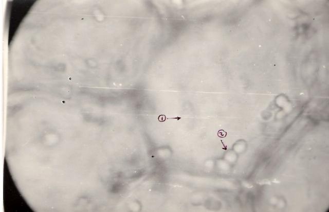
Cells from the pith of Vitis 1. Nucleus, with one nucleolus and irregular shape 2. Clusters of starch grains.
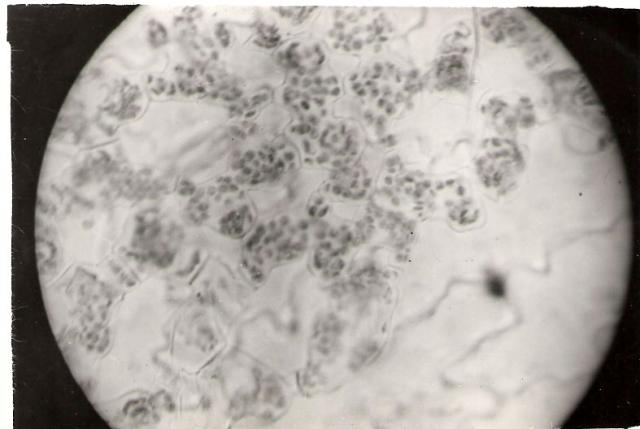
Chloroplasts of Viola tricolor leaf. 600X, 5 sec. Chloroplasts are little organelles inside cells that convert sunlight energy to sugars.
Mitosis in telophase. Onion root tip. Mitosis is the process of cell division.
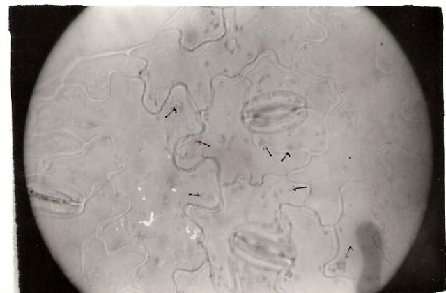
Stomata from Viola tricolor leaf. 600X, 5 sec. Stomata are gatekeepers on leaves who allow or disallow air to enter the plant tissue.

Post new comment

  • All spam and irrelevant comments will be deleted.
  • Comments posted here will take some time to appear on the site. Do not post your comment again if you do not see it. Just be patient and it will be published.
  • Note that what you post here will be publicly available on the web and will be indexed in search engines.
  • We reserve the right to unpublish any comments without stating the reasons for that.
  • All postings are subject to our Terms of use
The content of this field is kept private and will not be shown publicly.
  • Allowed HTML tags: <a> <em> <strong> <cite> <code> <ul> <ol> <li> <dl> <dt> <dd> <blockquote>
  • Lines and paragraphs break automatically.

More information about formatting options