Pollen grains often have very intricate shapes and structure. They have several layers, spines, pores, ...etc.
Pollen often has oils that protect it against dessication. These oils can hinder observing the structures, and may have to be washed in solvents.
|
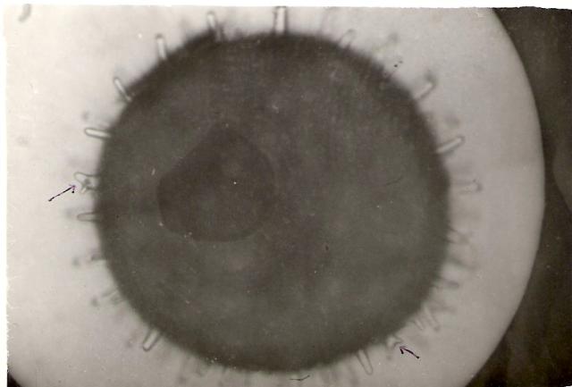
Hibiscus pollen. 600X, 5 sec. |
|
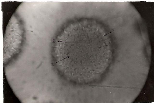
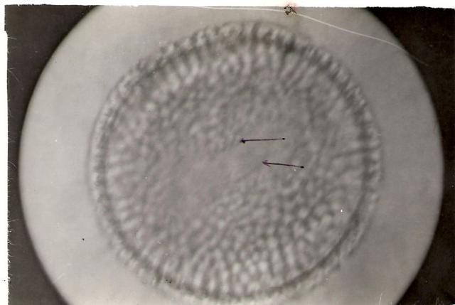
Pollen of Althea rosa. 600X, 5 sec. Note 1. spines and 2. pores |
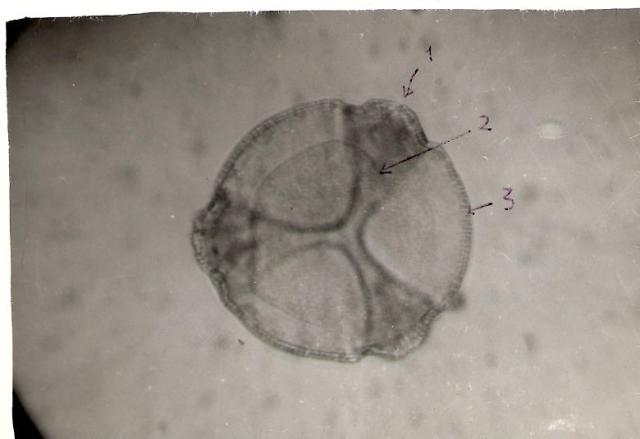 |
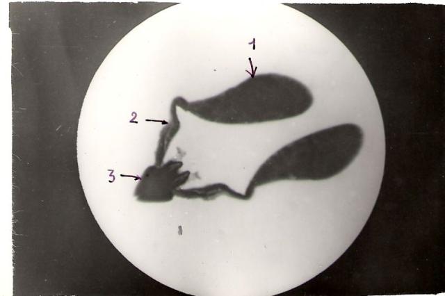
Pollen of Caesalpinia mounted in glycerin. 600X, 5 sec 1. Pore, 2. Furrow, filled with oily substance, 3. Baculate exine. |
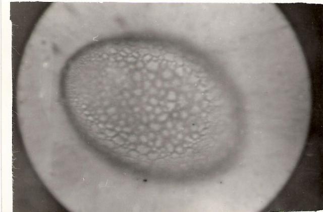 |
Pollen of Pancratium, in water. Note the reticulate bacula. 135X 15 sec |
 |
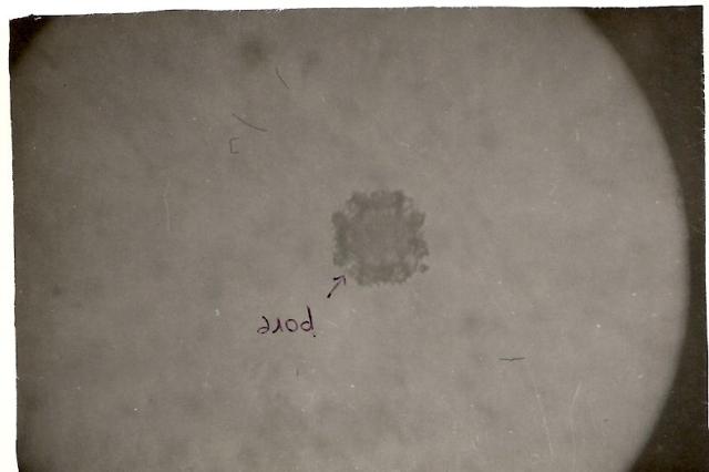
Pollen of Pelargonium, mounted in nail polish after cleaning in heptane. 1350X, 10 sec. Note: pore |
 |
Pollen of Sonchus (??) or related type. 600X, 5 sec. |
 |
Pollinium of Swamp Milkweed: "Asclepias curassavica" 1. Pollen Mass, 2. Translator, 3. Corpusculum. 56X, 1/5 sec. This is a very interesting adaptation. The entire pollen sac of the male flower is designed to cling to the pollinator insect (e.g. bee), and then delivered to the female flower. You can read more on the pollination process. |
Here are more sites where you can see many types of pollen at varying magnifications and techniques:
Contents:
Most Comments
Most commented on articles ...